Aert Medical
Magnetic Resonance Imaging (MRI) plays a crucial role in diagnosing joint conditions. Clinicians often face the question, "What is the difference between 0.5T and 1.5T MRI for joint imaging?". Understanding this difference is vital for optimal patient outcomes. A study from the Radiological Society of North America indicates that higher magnetic field strengths, like 1.5T, improve image resolution and detail.
The 1.5T MRI provides clearer images with better sensitivity for subtle injuries. A report by the American College of Radiology states that 1.5T MRIs can detect small joint effusions more effectively than their 0.5T counterparts. However, not all conditions require high-field MRI. The lower 0.5T MRI systems are often sufficient for routine evaluations and are cost-effective.
Each MRI type carries its own set of advantages and limitations. Some patients may also experience discomfort in higher magnetic fields. The decision should factor in the clinical scenario and patient preferences. As imaging technology evolves, the dialogue around the optimal choice continues to develop. We must remain attentive to these discussions, balancing technology's benefits against its practical applications.
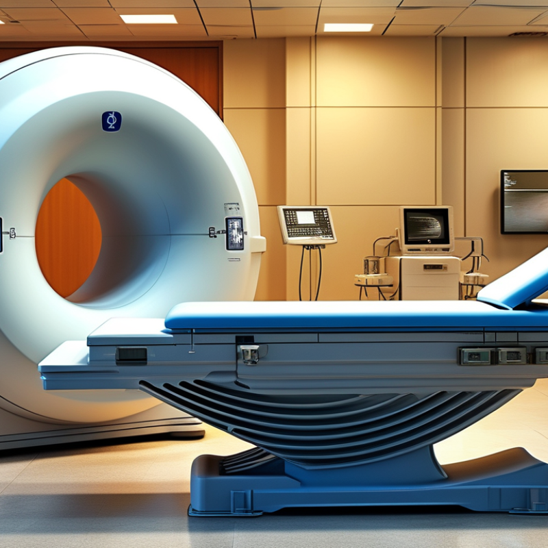
Magnetic resonance imaging (MRI) is crucial for assessing joint conditions. MRI technology operates with varying magnetic field strengths. The strength of the magnetic field is measured in teslas (T). Commonly, you will hear about 0.5T and 1.5T machines. Understanding these differences is vital for accurate diagnoses.
A 0.5T MRI provides lower-resolution images compared to a 1.5T MRI. This strength often results in longer scan times and less detailed results. However, it can be useful for general scans and for patients with mobility issues. These machines often have open designs, making the process more comfortable for some individuals. In contrast, 1.5T MRI machines produce clearer images with better contrast. The high field strength allows orthopedic specialists to detect subtle abnormalities in joint structures.
Still, it's essential to consider the practical aspects. Not every situation requires the highest strength. Factors such as patient comfort, cost, and specific clinical needs should guide the decision. Furthermore, some patients may experience anxiety in enclosed spaces during scans. Understanding these elements leads to better-informed choices.
When considering MRI for joint imaging, the choice between 0.5T and 1.5T systems significantly affects image quality. The strength of an MRI magnet is measured in Tesla (T). A 1.5T MRI is commonly used for detailed imaging due to its higher magnetic field strength. Research shows that 1.5T systems produce images with better resolution, allowing clearer visualization of soft tissues and cartilage structures. In joint imaging, this can be crucial for accurate diagnoses of pathologies.
Data indicates that 1.5T MRI can reduce motion artifacts by nearly 30% compared to 0.5T systems. This is essential for patients who may find it challenging to remain still. Additionally, studies have shown that 1.5T MRI can enhance the detection rate of subtle lesions, improving diagnostic accuracy. However, not all facilities have access to 1.5T machines, and 0.5T systems are still prevalent in many clinics. This presents a dilemma for practitioners when balancing image quality with availability.
Despite the advantages of 1.5T MRI, there are areas for further exploration. Notably, the differences in image quality may not be as pronounced in all patients or conditions. Some experts argue that certain joint issues can be effectively evaluated using 0.5T. This raises important questions about cost-effectiveness and the appropriateness of each MRI type for specific clinical scenarios. The choice should always be tailored to individual patient needs.
The bar chart above illustrates the average image resolution for 0.5T and 1.5T MRI scans. As shown, the 1.5T MRI offers higher resolution capabilities compared to the 0.5T MRI, leading to improved imaging quality, which is beneficial for joint imaging diagnostics.
When it comes to joint imaging, the choice between 0.5T and 1.5T MRI can significantly affect patient outcomes. A 0.5T MRI is often recommended for patients with certain conditions. This strength may be adequate for basic assessments. It is also typically more accessible and cost-effective. However, the image quality may not always meet the needs for detailed evaluation.
On the other hand, a 1.5T MRI provides higher resolution images. This strength is especially beneficial for diagnosing complex joint issues, such as tears or inflammation. The clarity offered by a 1.5T MRI can reveal details that a lower strength might miss. Yet, it also comes with higher costs and may not be necessary for straightforward cases. Each patient's condition must be critically assessed to choose the appropriate option.
Clinicians must weigh clinical necessity against available resources. In some instances, a 0.5T MRI could suffice, while in others, the superiority of a 1.5T could lead to better treatment decisions. The evolving technology in MRI machines prompts continuous evaluation of these choices. Personalizing each patient's imaging plan is essential for achieving optimal results.
When considering MRI options for joint imaging, cost is a crucial factor. The price difference between 0.5T and 1.5T MRI can be significant. A 0.5T MRI generally costs less upfront. This can make it appealing for facilities with tight budgets. However, lower initial costs do not always translate to long-term savings.
1.5T MRIs typically provide better image quality and detail. This can lead to more accurate diagnoses and reduce the chance of repeat scans. Facilities may save money by avoiding extra procedures. Patients may also experience better outcomes, which can lower overall healthcare costs.
Budgeting for MRI services requires careful thought. Some facilities might invest in 1.5T machines for their enhanced features. Yet, others may overlook the long-term benefits of image quality. In such scenarios, stakeholders must weigh immediate costs against potential future savings. Balancing economic factors with patient care can be a challenging endeavor.
| MRI Type | Magnetic Field Strength | Image Quality | Scan Time | Cost Per Scan | Availability |
|---|---|---|---|---|---|
| 0.5T MRI | 0.5 Tesla | Moderate | 30-45 minutes | $300-$500 | Common |
| 1.5T MRI | 1.5 Tesla | High | 15-30 minutes | $500-$900 | Widely Available |
When considering MRI scans for joint imaging, patient comfort and safety are crucial. The differences between
0.5T and 1.5T MRI machines extend beyond just imaging capabilities. Many patients express concern about how they
will feel during the procedure. For those who are claustrophobic, the 0.5T MRI can feel less confining. Its larger
openings may ease anxiety for some individuals.
However, comfort does not solely revolve around the machine's strength. The noise levels and the time taken during
the scan also play significant roles. A 1.5T MRI can provide clearer images, which may lead to a shorter scanning
time. Patients might prefer the speed but should be aware that this machine often has a smaller bore. This can
intensify feelings of confinement for sensitive individuals.
Safety remains a priority in both types of MRIs. While both machines are generally considered safe, patients should
disclose any concerns beforehand. Understanding potential risks, even in familiar settings, can promote trust
between patients and healthcare providers. Reflecting on personal experiences with MRI procedures may help guide
individuals in choosing the most suitable option for their needs.
: The main difference lies in the magnetic field strength. A 1.5T MRI provides higher image resolution than 0.5T.
It is recommended for basic assessments and certain conditions where high detail is not crucial.
A 1.5T MRI enhances detail in images. It helps diagnose complex joint issues better than a 0.5T MRI.
Not all facilities have access to 1.5T systems. Many clinics still use 0.5T machines.
Yes, in certain cases, it may provide adequate evaluation. It’s cost-effective and accessible.
A 1.5T MRI can reduce motion artifacts, making it beneficial for patients who can’t remain still.
Its higher costs may not justify its use for straightforward cases. Sometimes, a lower strength works just fine.
Personalizing imaging plans ensures the best outcomes for each patient based on their specific condition and needs.
Clinicians should weigh the clinical necessity against available resources, evaluating which MRI type is best suited.
Differences in image quality may not be significant for all patients or conditions. It’s an area needing further exploration.
The article titled "The Difference Between 0.5T and 1.5T MRI for Joint Imaging?" explores the key distinctions between two magnetic resonance imaging strengths commonly used for joint assessments. It begins with a foundational understanding of MRI technology, emphasizing how magnetic field strength affects imaging capabilities. The article highlights that higher field strengths, such as 1.5T, generally produce superior image quality, allowing for more detailed visualization of joints, which is crucial for accurate diagnosis.
In discussing clinical implications, the article addresses scenarios where a 0.5T MRI may be preferred, such as when patient safety and comfort are priorities or when cost considerations are significant. The economic aspects of MRI options are examined, noting that 0.5T MRIs are often more cost-effective, while 1.5T MRIs provide advanced imaging features that may justify their higher price in certain situations. Ultimately, the article answers the question, "What is the difference between 0.5T and 1.5T MRI for joint imaging?" by balancing the benefits of image quality and patient experience against economic factors.