Aert Medical
Why are 0.5T MRI systems more cost-effective for private practices? The answer begins with practical economics, not marketing claims. A 0.5T scanner usually requires lower capital investment, simpler site preparation, and reduced cooling demands than many high-field systems. These differences can matter when a clinic has limited space, modest patient volume, or strict financing limits.
Industry evidence supports a careful comparison. The OECD Health Statistics database shows substantial differences in MRI availability and utilization across healthcare systems, reminding operators that purchasing decisions depend on local demand. The IMV 2023 MRI Market Summary Report also emphasizes equipment utilization, service costs, and workflow efficiency as important ownership factors. Meanwhile, Grand View Research has projected continued growth in the global MRI market, reflecting rising diagnostic demand and stronger competition among outpatient providers.
Radiologist and low-field MRI researcher Dr. Andrew Webb has stated, “Low-field MRI is not just a cheaper version of high-field MRI.” That distinction matters. A 0.5T system may serve musculoskeletal, neurological, abdominal, and routine follow-up examinations, but its clinical limits must be assessed honestly. Some difficult studies may still require 1.5T or 3T imaging.
For a private practice, the financial picture becomes clearer when the scanner is busy, the room is compact, and maintenance interruptions are limited. Lower operating pressure can also support a calmer patient experience. Still, cost-effectiveness is not automatic. Referral patterns, reimbursement, radiologist expertise, installation costs, and local regulations can change the result. A spreadsheet may look convincing. The real test is daily workflow.
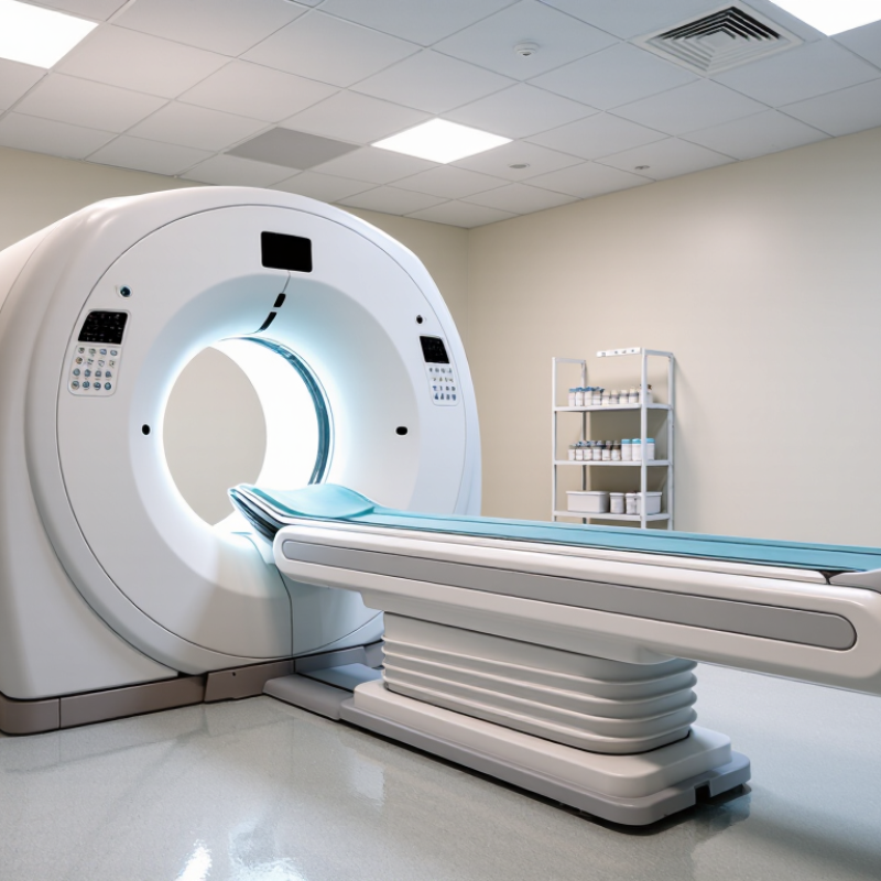
A 0.5T MRI system uses a magnetic field of 0.5 tesla. That is lower than most hospital systems. Yet it can still produce useful images for joints, the spine, the brain, and routine musculoskeletal examinations.
Many 0.5T systems use permanent magnets. They usually need less complex cooling infrastructure. This may reduce room preparation, service demands, and installation costs for private practices. Some designs also provide a more open patient space. That matters when a patient feels anxious inside a narrow scanner.
Clinical performance still depends on protocols, coils, software, and operator experience. Lower field strength can mean weaker signal and longer examinations. That trade-off is easy to underestimate. A 2023 review in European Radiology reported growing interest in low-field MRI because modern reconstruction methods can recover useful diagnostic detail. However, advanced imaging remains more demanding.
The economic case is practical. The OECD Health Statistics 2023 dataset shows wide differences in MRI availability between countries, suggesting uneven access and local demand. A smaller practice can place a 0.5T unit closer to patients instead of sending every referral elsewhere. Grand View Research valued the global MRI market at approximately 7.1 billion dollars in 2023, reflecting continued investment in outpatient imaging.
Operators should compare scan time, referral mix, maintenance, staffing, and reimbursement. The cheapest scanner is not always the most profitable one. That lesson deserves a second look.
A 0.5T MRI uses a 0.5-tesla static magnetic field to align hydrogen protons in the body. That equals approximately 5,000 gauss. A radiofrequency pulse then disturbs this alignment. As protons recover, they release signals that form tissue images. At 0.5T, the proton resonance frequency is about 21.3 MHz, calculated from the accepted 42.58 MHz-per-tesla relationship.
The process is gentler in several practical ways. Lower field strength generally reduces susceptibility artifacts and can improve imaging near air, metal, or bone. The FDA confirms that MRI uses non-ionizing energy, unlike X-ray-based imaging. However, lower field strength also produces less signal. Some examinations may require longer acquisition times or stronger signal-averaging methods. The compromise is real. A small clinic should not assume every study will be faster.
Safety still depends on operating conditions, not field strength alone. The ACR Manual on MR Safety emphasizes screening, controlled access, and documented implant checks. IEC 60601-2-33 specifies whole-body SAR limits, including 2 W/kg in Normal Operating Mode. A 0.5T system may simplify siting and reduce infrastructure demands, supporting private-practice economics. Yet workflow matters more than magnet strength alone. Patient positioning, coil selection, and technologist experience directly influence usable image quality. A 2023 outpatient imaging market report from IMV also identifies throughput and utilization as major cost drivers. That finding deserves attention. A lower-cost scanner still becomes expensive when appointments remain empty.
A 0.5T MRI can support private practices that need practical imaging without purchasing a high-field system. Its lower field strength may create a calmer scanning environment, with less noise and a more open patient experience. This can help anxious adults, older patients, and some children remain still during examinations.
Which clinical applications suit it best? Musculoskeletal imaging is a strong fit. Practices can assess knees, shoulders, wrists, ankles, and soft-tissue injuries with dedicated positioning and focused protocols. Spine examinations may also be appropriate for disc disease, stenosis, and follow-up imaging. Brain studies can support selected evaluations, including routine neurological assessment and monitoring known conditions.
Abdominal and pelvic imaging require more careful planning. Motion, body habitus, and lower signal can affect image quality, so experienced technologists and optimized coils matter. Scan times may be longer. That matters in busy clinics. Some urgent or highly detailed studies may still favor higher-field equipment, depending on local protocols and radiologist requirements. The system is not a universal replacement. It is a clinical tool with boundaries. Private practices should review referral patterns, reporting expertise, patient volume, and accreditation standards before choosing applications. A small clinic may gain value from dependable routine studies, but occasional difficult cases can expose limitations that are easy to underestimate.
| Clinical Application | Typical Use | Suitability at 0.5T | Key Considerations | Practice-Level Value |
|---|---|---|---|---|
| Musculoskeletal imaging | Assessment of many routine joint, tendon, ligament, and soft-tissue complaints, such as knee or shoulder pain. | Often suitable when the protocol and image quality meet the diagnostic question. | Lower field strength can mean lower signal-to-noise ratio and longer scan times than higher-field systems. Small structures and subtle abnormalities may be more challenging. | Can support a focused outpatient service with routine referrals and appropriate radiologist review. |
| Spine imaging | Evaluation of common degenerative conditions, disc disease, and selected spinal canal or nerve-root concerns. | Often suitable for routine examinations, depending on patient size, motion, and the clinical question. | Protocol selection and adequate image quality are essential. Complex cases or subtle findings may require referral for higher-field imaging or another modality. | May serve a steady outpatient demand without requiring every case to be performed on a high-field scanner. |
| Brain imaging | Selected routine examinations, including assessment of some structural or chronic neurological conditions. | Case dependent; suitability varies with the indication and required sequences. | Some small lesions, subtle abnormalities, and advanced applications may be less well assessed than on higher-field systems. Follow established protocols and referral pathways. | Can broaden access for appropriate examinations, while complex neurological studies may be directed to a higher-field service. |
| Abdominal and pelvic imaging | Selected examinations of abdominal or pelvic organs when local protocols and clinical requirements are appropriate. | Selective; not every abdominal or pelvic indication is a good fit. | Motion, breath-holding, field of view, and signal limitations can affect image quality. Some detailed or time-sensitive studies may be better suited to other equipment or modalities. | Best considered as a targeted service after reviewing local referral patterns and the capabilities of the specific system. |
| Cardiac imaging | Specialized assessment of cardiac structure, function, or tissue characteristics. | Generally limited compared with systems and services specifically configured for cardiac MRI. | Cardiac MRI requires specialized hardware, sequences, expertise, and workflow. Confirm system capability and clinical validation before offering these examinations. | Usually not a core economic justification for a general-purpose 0.5T installation. |
| Vascular imaging | Selected vascular examinations, depending on anatomy, protocol, and whether contrast is required. | Selective; feasibility depends on the system and the diagnostic task. | Flow effects, spatial resolution, scan time, and contrast requirements matter. Confirm the examination is supported by the scanner and local protocols. | Potentially useful for a defined referral niche, but should not be assumed to replace higher-field vascular imaging. |
| Claustrophobia-sensitive patients | Examinations for patients who may tolerate an open or more spacious scanner design better. | Potential operational advantage when the scanner design improves patient acceptance. | “Open” describes scanner design, not guaranteed comfort or suitability. Patient tolerance varies, and image quality depends on the specific system and protocol. | May reduce some appointment barriers and support patient access; assess acceptance and completion rates locally. |
| Capital and operating costs | Equipment purchase, room preparation, staffing, service, utilities, and ongoing quality assurance. | Potentially favorable, not guaranteed relative to higher-field options. | Actual total cost depends on the scanner, installation, financing, service agreement, staffing, local regulations, and throughput. Lower field strength alone does not establish lower lifetime cost. | Model total cost of ownership against realistic scan volume, reimbursement, exam mix, and expected appointment times. |
A 0.5T MRI can fit private practice budgets more comfortably than higher-field systems. Its value comes from total ownership, not purchase price alone. Lower-field equipment may require fewer electrical upgrades and simpler room preparation. Smaller site requirements can also reduce construction costs. These savings matter when a clinic works within a fixed renovation budget.
Energy use, cooling needs, and service requirements can influence monthly expenses. A practice may also need fewer infrastructure changes before installation. Staff training remains essential, especially for protocol selection and patient positioning. Experienced technologists can help produce consistent examinations. Radiologists must also understand the system’s clinical strengths and limitations.
The calculation is not always neat. A lower-field scanner may provide useful studies for joints, routine neurological work, and selected musculoskeletal examinations. However, some scans may take longer or show less detail than higher-field imaging. That difference can affect scheduling and patient satisfaction. A cheaper scanner can become expensive if referrals require frequent repeat imaging. Utilization should be estimated from local demand, reimbursement, staffing, and expected scan volume. Maintenance contracts, accreditation, safety procedures, and room downtime also deserve careful review. Clinical fit matters more than attractive numbers on a quotation.
Why Is a 0.5T MRI Cost Effective for Private Practices?
What Should Private Practices Consider Before Adoption?
A 0.5T MRI may reduce purchase, installation, and shielding expenses. It can also fit smaller outpatient sites with fewer construction changes. However, lower field strength does not guarantee lower total cost. OECD Health Statistics 2023 shows wide differences in MRI availability, ranging from fewer than 10 to more than 50 units per million people. Local demand matters more than a global average.
Private practices should assess referral volume, examination types, staffing, and service contracts. A 0.5T system may support routine musculoskeletal, neurological, and spine examinations. Complex vascular or advanced body imaging may require longer protocols or additional review. The American College of Radiology’s MR Accreditation Program emphasizes image quality, safety procedures, qualified personnel, and quality control. These requirements still apply when the magnet is smaller.
Workflow deserves close attention. A slower examination can reduce daily capacity, even when the scanner costs less. Clinics should model appointment length, repeat-scan risk, maintenance downtime, and patient no-shows. A simple spreadsheet is useful, but it can miss referral changes. That is the uncomfortable part. Cost projections often look precise while relying on uncertain assumptions. Practices should test demand with local referral data and review outcomes after six months. Patient comfort, room design, emergency access, and staff training also influence adoption. The cheapest installation may become expensive if it creates delays, repeat imaging, or limited clinical use.
A 0.5T MRI system can reduce capital, service, and facility costs, but the financial advantage depends on examination volume, scan time, staffing, reimbursement, and clinical scope. The chart below presents a transparent planning model rather than a supplier quotation.
Planning assumptions: 7-year equipment depreciation, 1,500 examinations per year, estimated equipment investment of $500,000 for 0.5T and $1,200,000 for 1.5T, annual service costs of $50,000 and $100,000, staffing costs of $150,000 for each system, and annual facility and utilities costs of $15,000 and $25,000. Local quotations, staffing models, reimbursement, and patient demand should be validated before adoption.
It describes the magnet’s field strength: 0.5 tesla, or about 5,000 gauss. The number alone does not determine image quality.
It can produce useful images of joints, the spine, the brain, and routine musculoskeletal conditions. Suitability depends on the examination and clinical needs.
The magnetic field aligns hydrogen protons in the body. A radiofrequency pulse disturbs them, and returning signals help form tissue images. The process uses non-ionizing energy.
Yes. Lower field strength can mean less signal, longer scans, or added signal averaging. The trade-off is real.
Some designs offer a more open patient space than narrow scanners. That may ease anxiety, though the room and examination can still feel unfamiliar.
Some systems use permanent magnets and need less complex cooling. They may also require fewer room changes or electrical upgrades. Actual costs vary.
Estimate local demand, staffing, scan volume, reimbursement, maintenance, and downtime. Include training and room preparation. A low purchase price can be misleading.
Choose suitable protocols and coils, position each patient carefully, and use experienced staff. These details matter. I may be oversimplifying, but magnet strength alone is not the whole story.
A 0.5T MRI system is a low- to mid-field magnetic resonance imaging solution that uses a 0.5-tesla magnetic field to produce diagnostic images without relying on the higher operating costs of stronger systems. Its technology is well suited to routine examinations, including musculoskeletal, neurological, spinal, and selected abdominal or pelvic studies, depending on image-quality requirements and clinical protocols. Although it may offer lower signal strength than high-field MRI, modern software, optimized coils, and efficient scanning workflows can support dependable everyday imaging.
Why are 0.5T MRI systems more cost-effective for private practices? They generally require lower installation, infrastructure, energy, and maintenance investments, while their compact design can simplify site preparation and improve room utilization. Before adoption, a practice should assess patient volume, referral patterns, available space, staffing, expected scan times, image-quality expectations, regulatory requirements, and long-term service support. When matched with appropriate clinical needs, a 0.5T MRI can expand imaging capabilities while helping control capital and operating expenses.